Early Detection
Breast Biopsies
What is a breast biopsy?
A breast biopsy is a process in which cells or small pieces of breast tissue are removed from an area of concern in the breast such as a lump, changes on the mammogram or the ultrasound. (Ref 1-3).
Following completion of the biopsy a small metal marker, referred to as a “clip” or a “tissue marker”, is placed in the biopsy site so that future imaging can identify the area of the previous biopsy.
The biopsy specimen is sent to the pathologist to evaluate whether the removed specimen is benign or malignant.
In some cases, the initial biopsy will be non-diagnostic and a repeat biopsy may be required. In other cases, the biopsy will be classified as high-risk (i.e., LCIS or atypia). In both cases the patient would be referred to a breast surgeon.
There are three types of biopsies:
Core needle biopsy
In this procedure a needle approximately the size of the lead in a pencil is directed to the area of concern and “pieces” of tissue are removed and sent to the pathologist. The core needle biopsy procedure is the preferred method of determining if a lump or change on breast imaging is benign or malignant.
Fine needle aspiration
Fine Needle Aspiration is an important, but less commonly performed procedure, in which a “small needle” is placed in the area of concern and a sample of “cells” is removed. We typically reserve this procedure for women who present with an obvious advanced breast cancer. This procedure can provide a diagnosis of cancer within hours and expedites treatment planning.
Open surgical biopsy
Open surgical biopsy is a procedure in which a surgeon makes an incision in the breast and removes the entire area of clinical concern. To remove the appropriate amount of tissue, the radiologist often places one or more wires in the breast prior to the surgical procedure to assist the surgeon in identifying and removing the tissue of concern. This procedure is referred to as “wire localization”.
Techniques used in making the diagnosis
Stereotactic core biopsy
In this procedure the patient lies face down on a table. There is an opening in the table that allows the breast to be positioned below it. A series of mammograms are taken so that a 3D image can be created by the computer to precisely locate the area of concern. A small incision is made, the needle is then guided to the exact area of concern and multiple biopsies are taken.
Ultrasound guided core biopsy
If the area of concern can be seen on the ultrasound, an ultrasound guided biopsy is usually recommended as the procedure of choice. This procedure is typically better tolerated and quicker to perform than a stereotactic biopsy.
MRI guided biopsy
This procedure is limited to cases in which the area of concern is only seen on the MRI. (Ref. 4)
Nipple discharge
In cases of spontaneous nipple discharge that is either clear or blood tinged, a surgical biopsy is required to make the diagnosis of what is causing the nipple discharge. A mammogram should be performed before the surgical biopsy to determine if there are any changes in the breast that might be identified that could be causing the discharge.
If an area of concern is noted, the radiologist will assist the surgeon by placing a localization wire next to the area of concern. More commonly the mammogram is negative, and the radiologists inject the draining duct with a blue dye which makes it easier for the surgeon to accurately detect the proper duct in question for the biopsy.
In cases in which neither the mammogram nor the blue dye identifies an area of concern the surgeon will be required to remove the entire duct in the operating room from just below the nipple to the muscles below (Ref. 5).
In most cases the biopsy will be a benign growth commonly referred to as papilloma. In the unusual case in which a cancer is detected, options for additional surgery will be discussed with the patient.
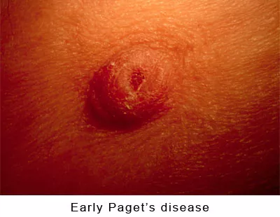
Paget’s disease of the Nipple
Paget’s disease of the nipple is a clinical condition that is identified by skin changes in the nipple. The diagnosis is often missed in its early stage in which it often looks like eczema or non-specific inflammation. See pictures below of an early Paget’s disease. Any question about skin changes in the nipple should be brought to the attention of a breast surgeon. The condition is often misdiagnosed by dermatologists.
The diagnosis of Paget’s disease can be made in the physician’s office using local anesthesia. Under local anesthesia the surgeon uses a small circular blade to remove a sample of the area of concern. The procedure, Punch Biopsy, takes about 20 minutes and is very well tolerated (Ref 6).
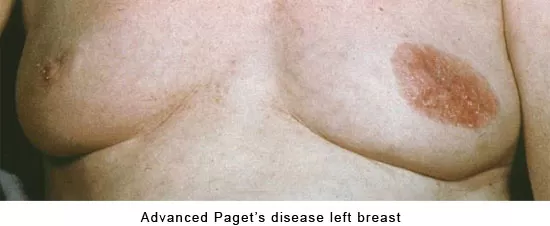
Below pictures of early Paget’s Disease and Advanced Paget’s Disease
Donate
Today
Your donation supports our mission to provide breast cancer education to EVERY woman.